

Click image to enlarge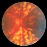 Fig. 8 Argon laser panretinal photocoagulation for the treatment of proliferative diabetic retinopathy. |
Photocoagulation of the Posterior Segment
Using topical anesthesia and a contact lens, the laser light for photocoagulation can be delivered into the eye by a slit-lamp delivery system. Diseases or conditions which are treatable by photocoagulation include diabetic proliferative retinopathy (fig. 8), diabetic macular edema, branch retinal vein occlusion, peripheral retinal and choroidal neovascularization, idiopathic central serous chorioretinopathy, vascular anomalies, nonvascular tumors, and retinal breaks. The spot size is dependent on the location of the treatment: 50–200 µm for macular photocoagulation, 200–1,000 µm for peripheral retinal spots. Power and burn duration are initially set according to desired burn intensity, and the aiming beam intensity is set to the lowest level that permits adequate beam visualization. The retinal burn, or opacification, produced during photocoagulation is due to protein denaturation in the outer retina [5].
Photodynamic Therapy
Age-related macular degeneration is a major cause of severe vision loss in people older than 65 years in North America and Europe. Loss of visual acuity results from choroidal neovascularization (CNV). The standard treatment for choroidal membranes involves photocoagulation with a hot laser, which inevitably destroys the adjacent and overlying normal retina. Now a new treatment method for CNV is available – photodynamic therapy with verteporfin (VisudyneTM) – that can minimize damage to the surrounding viable retinal tissue. In this treatment, 15 min after the verteporfin has been injected intravenously, the drug is activated by delivering a cold laser light (at 689 nm) over 80 s, using a spot size with a diameter 1,000 µm larger than the greatest linear dimension of the CNV lesion. A recent comparative, placebo-controlled study has recommended verteporfin therapy for the treatment of patients with predominately classic CNV resulting from age-related macular degeneration [6].
Conclusion
Lasers have brought about a revolution in ophthalmic surgery: Owing to their precision and noninvasive nature, the actual surgery can be performed on an outpatient basis in a matter of minutes, with little or no pain or discomfort. Recovery time after treatment is short as there are no large incisions to heal and postoperative complications, e.g. inflammation and infection, are reduced substantially. Today, almost 40 years after the advent of the laser, medical laser technology continues to evolve rapidly and ophthalmologists continue to explore new applications for this tool, whose possibilities for saving and sharpening vision seem to be unlimited.
The Beginnings
How Do Lasers Work?
Anterior Segment Surgery
Refractive Surgery
Pediatric Eye Surgery
Retinal Surgery
Conclusion
References
Biography
>> next