The skin can be divided into three layers: the external epidermis, the dermis below, itself subdivided into two layers, and the subcutis.
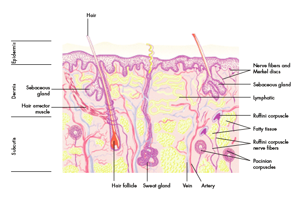
The epidermis varies in thickness between 0.1 and 1 mm. It consists primarily of epithelial cells – mainly keratinocytes which are increasingly differentiated as they migrate from the basement membrane (separating dermis and epidermis) to replace lost cells, a process taking 2–3 weeks. Melanocytes are also found in the basal layer of the epidermis. The epidermis has no blood vessels and must receive nutrients via diffusion from the dermis. The keratin in the outer epidermal cells forms an effective barrier against desiccation and bacterial and toxin entry. It is also a neurosensory and social-interactive surface.
The dermis is a dynamic layer of thick connective tissue, also in constant turnover. The elasticity and durability of the dermis provide the body protection against trauma. The thinner outer papillary dermis contains capillaries, elastic and reticular fibers and some collagen. The connective tissue in the reticular dermis is thicker; it contains larger blood vessels, elastic fibers and collagen. The primary dermal cell is the fibroblast, which produces collagen, elastin and the extracellular matrix of mucopolysaccharides, chondroitin sulfate and glycoproteins. Mast cells are also present as are most nerve endings (e.g. the pressure- and temperature-sensitive Ruffini corpuscles, and the pressure-sensitive pacinian corpuscles), lymphatics and skin appendages.
The subcutis is essentially a layer of fatty tissue containing vascular and neural structures. It serves for insulation and energy storage.
Important skin appendages are: sebaceous glands which secrete sebum to lubricate the skin and make it more impermeable to moisture; sweat glands producing sweat to cool the body by evaporation; apocrine glands which produce odor, and hair follicles. All these appendages are lined with epithelial cells which can be recruited for repair when the epithelium is damaged.
Skin is thickest on the palms and soles of the feet, thinnest on the eyelids and behind the ear. Male skin is characteristically thicker than female's and children's thin skin thickens progressively until the 4th or 5th decades of life when it begins to atrophy with loss of elastic fibers, skin appendages and matrix.
Figure adapted from: G. Thews, E. Mutschler, P. Vaupel: Anatomie, Physiologie und Pathophysiologie des Menschen. Stuttgart, Wissenschaftliche Verlagsgesellschaft, 1999
|